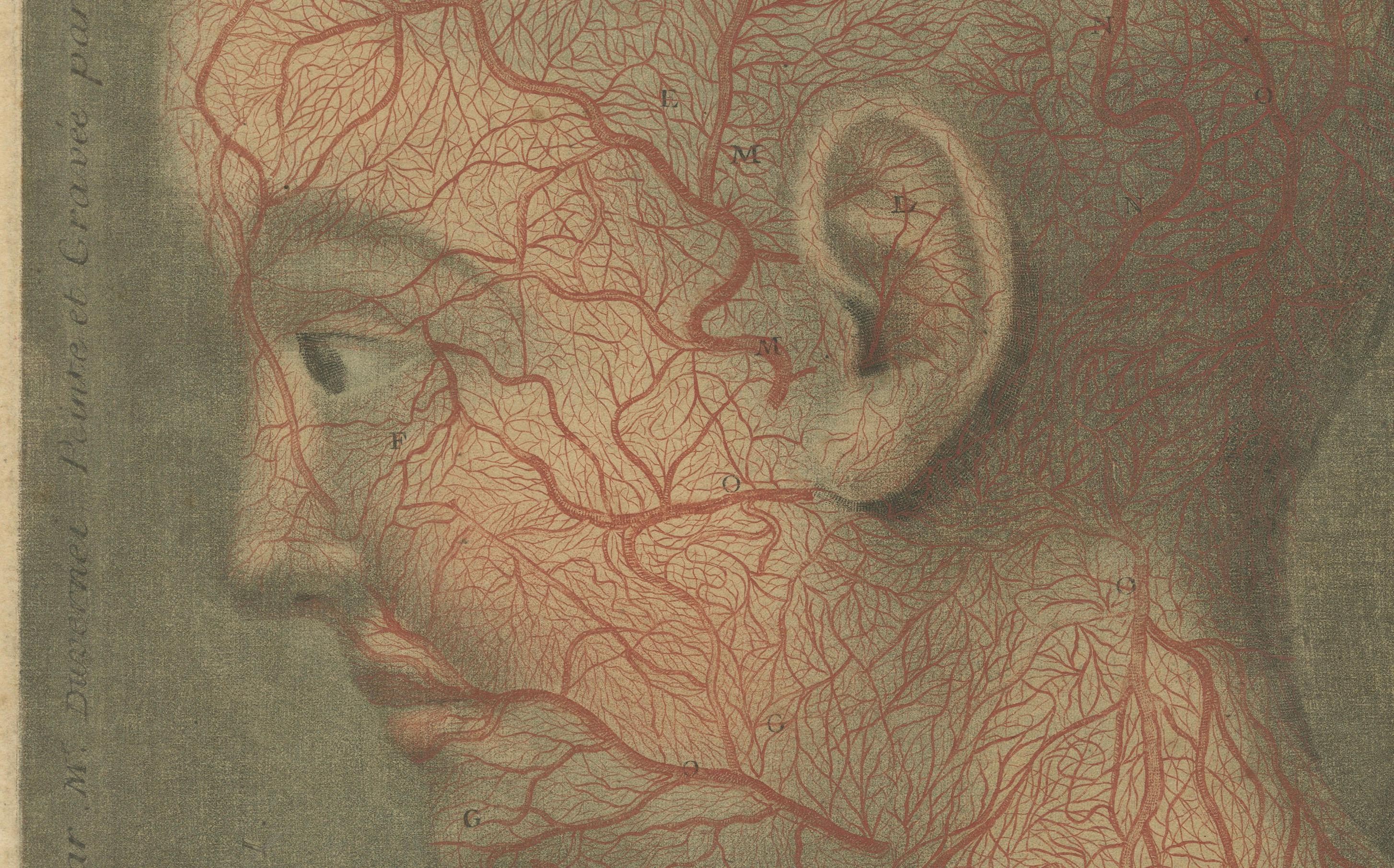
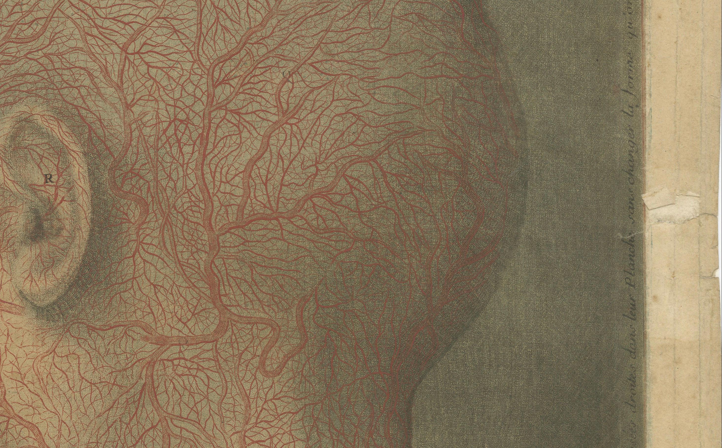
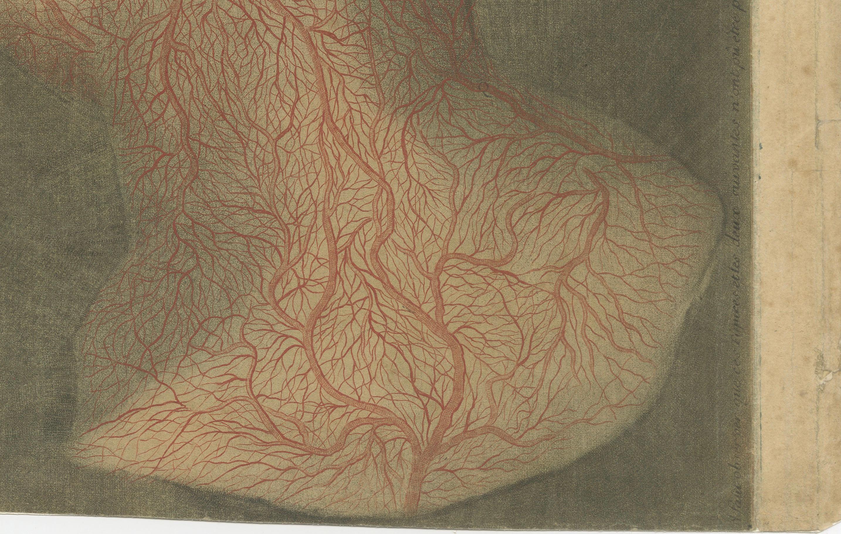
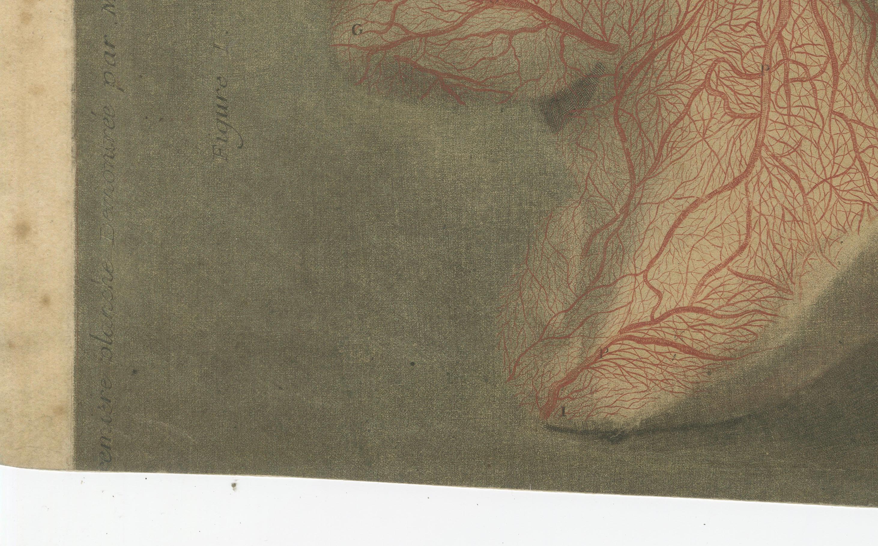
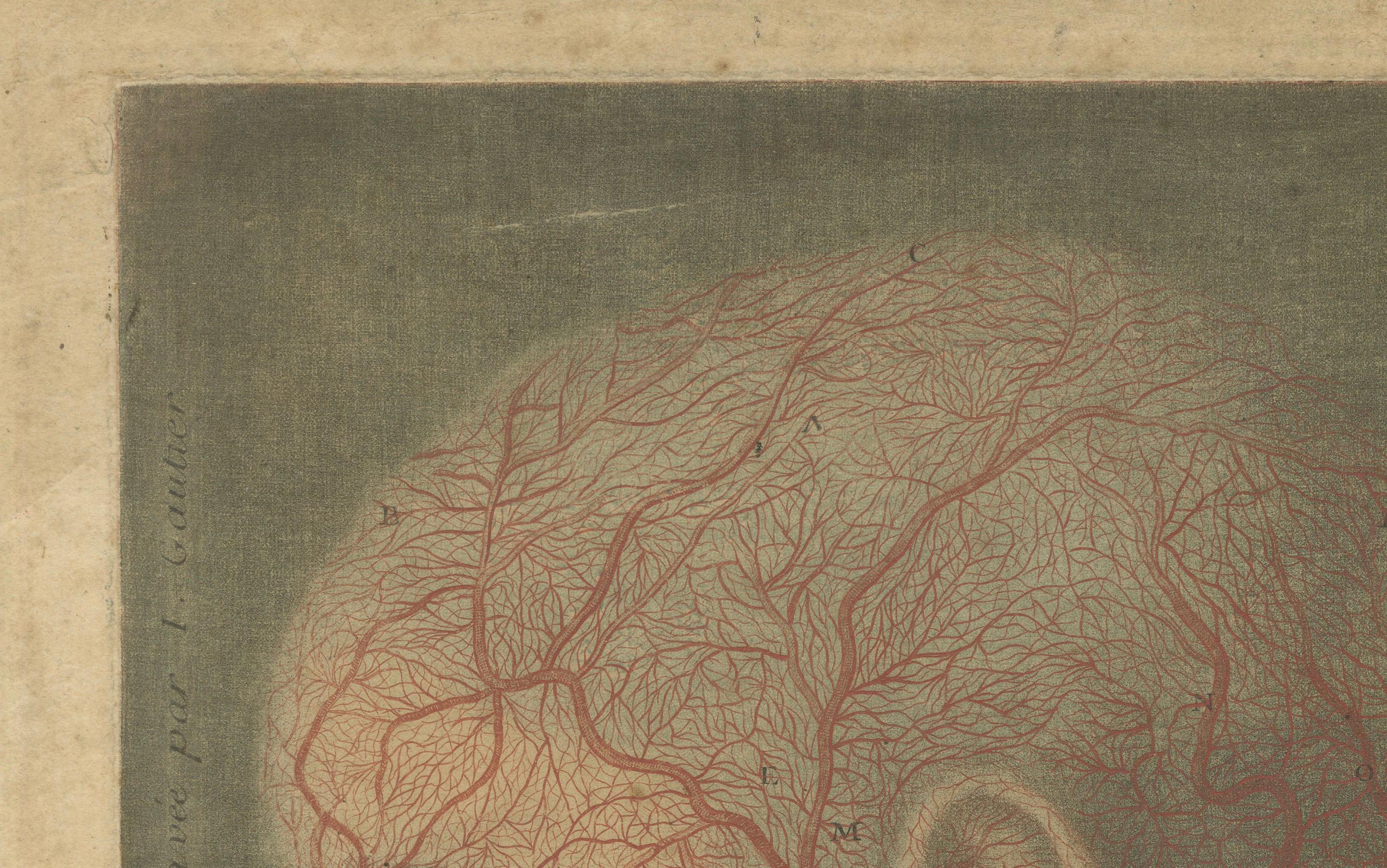
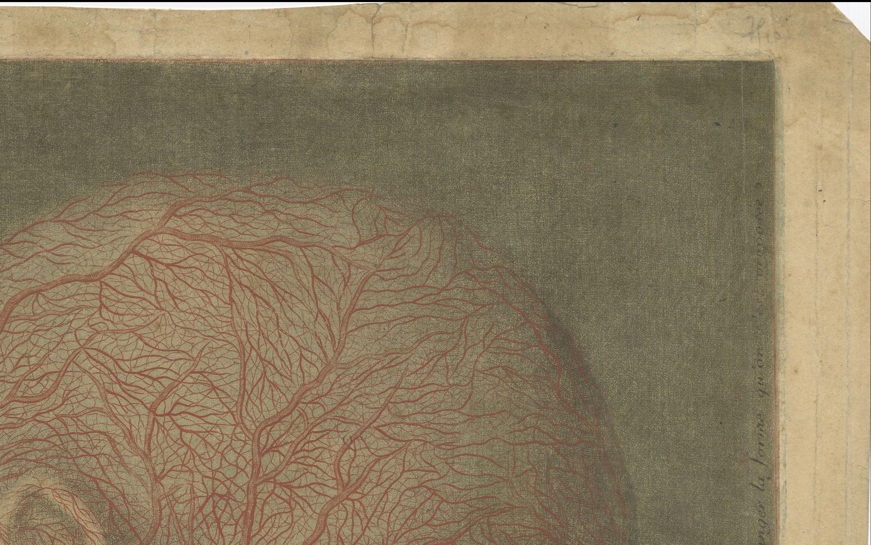
Anatomical Study of Head and Neck Veins – Rare Color Mezzotint, 1748
€ 990
One of a kind: this is the sheet in the photographs. Ships from Langweer, the Netherlands.
Date
1748
Sheet size
31.5 × 40 cm (12.4 × 15.7 in)
Condition
Fair
Reference
BG-14096
Condition
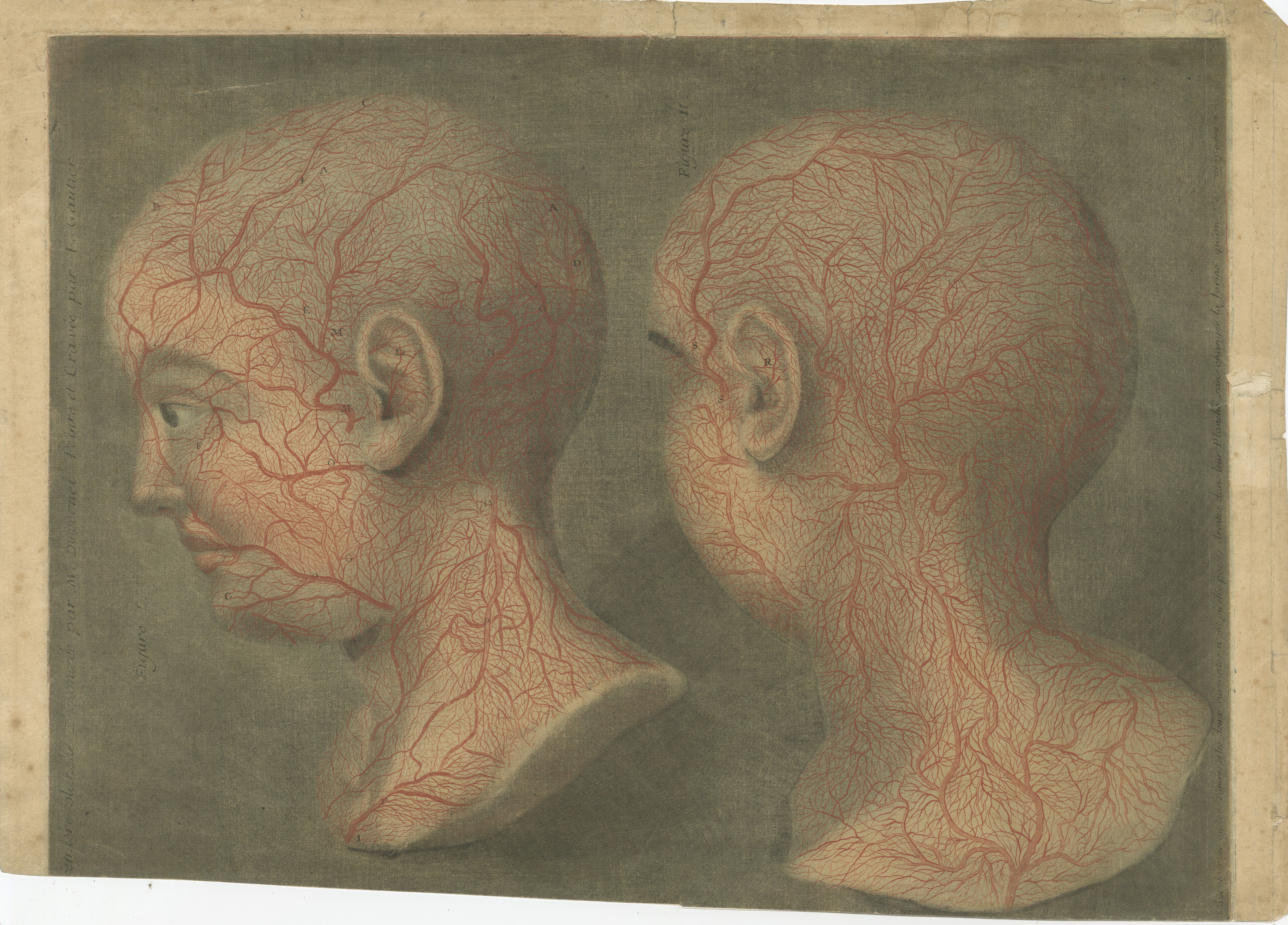
Margins trimmed up to 1.5 cm, with a small tear at right touching the image. Overall well preserved and visually impactful. A rare and attractive example of early color anatomical printing.
About this sheet
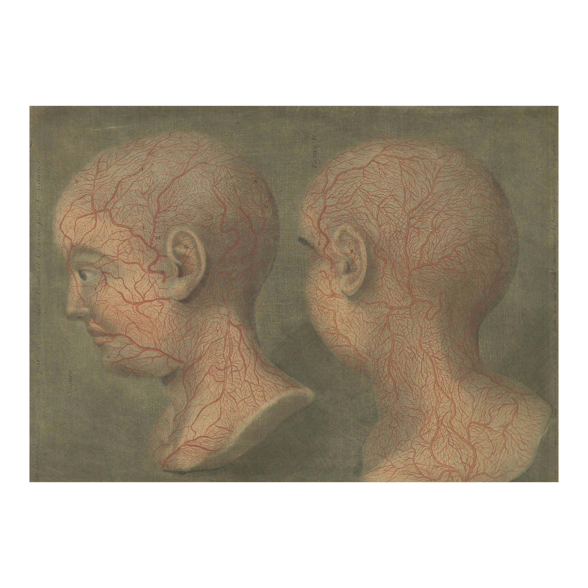
Anatomical Study of Head and Neck Veins – Color Mezzotint, 1748
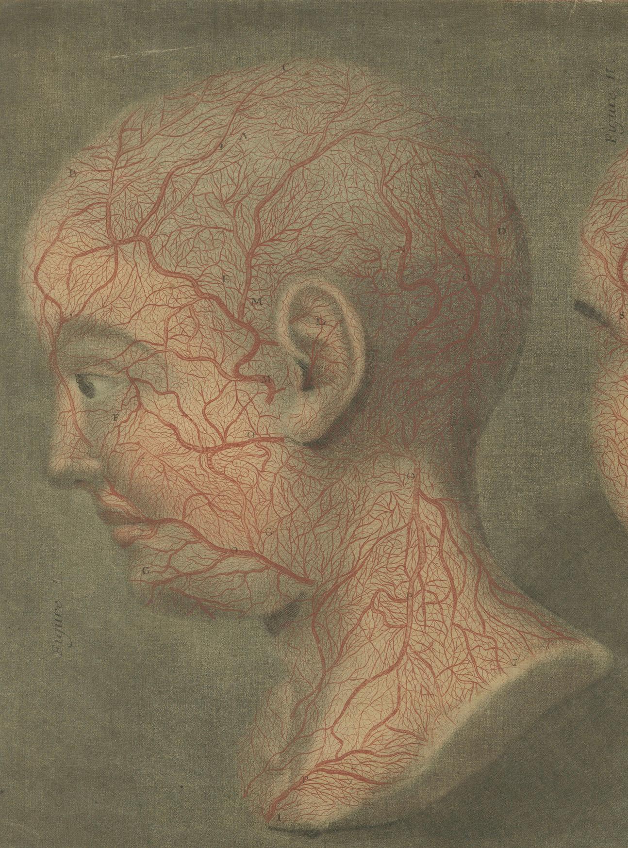
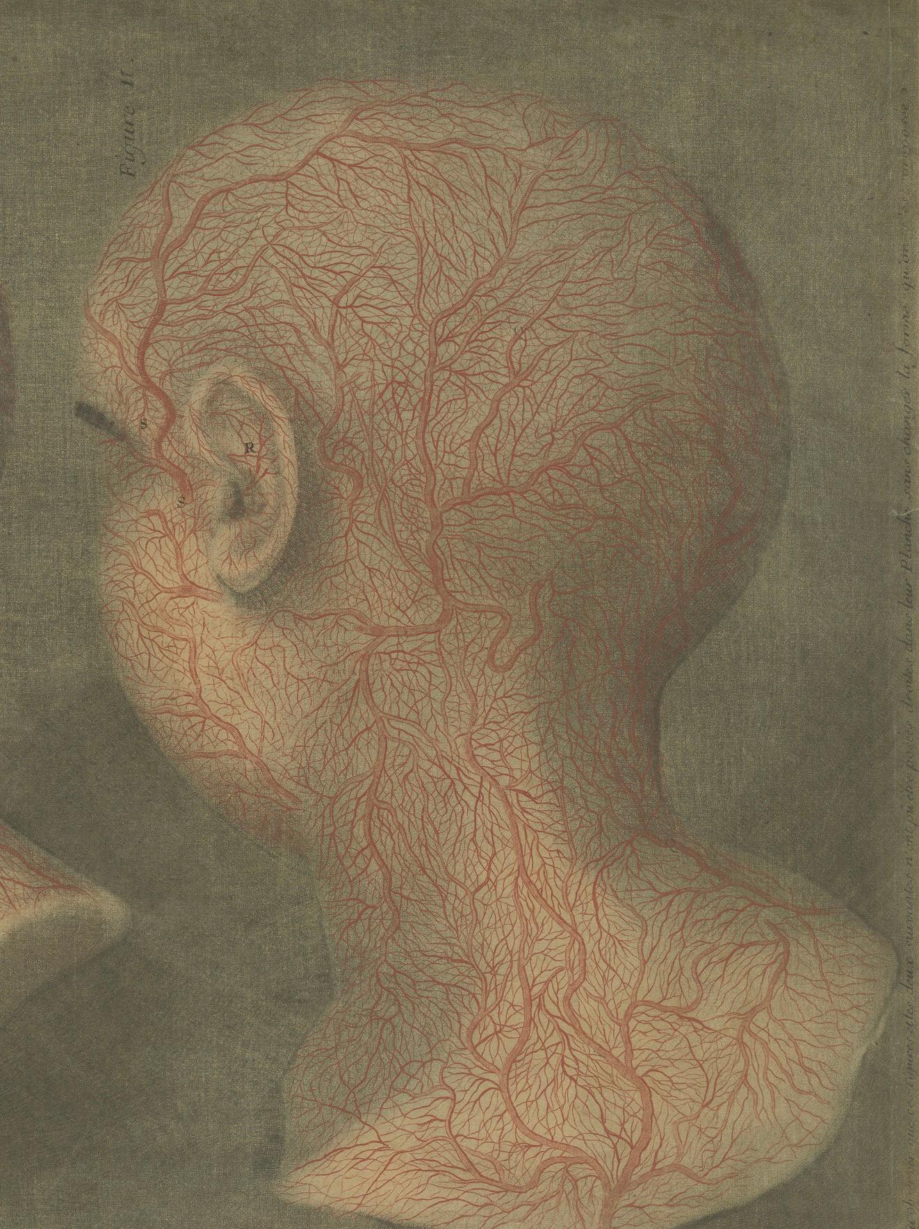
This striking 18th-century anatomical print, created by Jacques-Fabien Gautier-D’Agoty (1710–1781), presents a meticulous depiction of the superficial blood vessels of the human head and neck. Rendered using the color mezzotint technique after preparations by anatomist Joseph Guichard Duverney, the composition shows two views: the left profile and rear of a human head, each delicately traced with the extensive network of veins and arteries in vivid red against a soft neutral background.
This engraving was originally published in Gautier-D’Agoty’s landmark anatomical folio 'Anatomie de la tête, en tableaux imprimés', issued in Paris in 1748. Gautier-D’Agoty was a French anatomist, printmaker, and painter known for his pioneering use of color in mezzotint printmaking—a technically demanding process rarely employed for medical illustration before his time. His collaboration with Duverney, a respected anatomist of the Paris Academy of Sciences, produced a series of anatomical plates that were groundbreaking not only for their scientific accuracy but for their almost surreal visual presence. Garrison-Morton recognized the series as “remarkable for its striking mezzotints printed in color.”
This plate exemplifies the fusion of art and science in Enlightenment France. The use of layered color printing gives the circulatory structures an almost lifelike presence. The viewer is not only informed but also aesthetically moved by the eerie calm of the expressionless face and the fine tracery of the vascular system.
Such prints are extremely rare and highly sought after by collectors of medical history, early printing, and Enlightenment-era scientific illustration. They stand at the crossroads of knowledge and beauty, encapsulating a pivotal moment in the history of anatomical education.
Condition summary: Margins trimmed up to 1.5 cm, with a small tear at right touching the image. Overall well preserved and visually impactful. A rare and attractive example of early color anatomical printing.
Framing tips: Frame in black or dark walnut with an archival mat to highlight the red tones. Use non-reflective glass to reduce glare and preserve detail. Ideal for study, office, or gallery.