One of a kind: this is the sheet in the photographs. Ships from Langweer, the Netherlands.
Date
1867
Sheet size
28.5 × 22 cm (11.2 × 8.7 in)
Condition
Good
Reference
BG-12980-2
Condition
Good condition for its age, with light toning and scattered small foxing spots mainly in the margins. Paper remains strong with no tears or repairs, and the engraving lines are crisp and well-preserved.
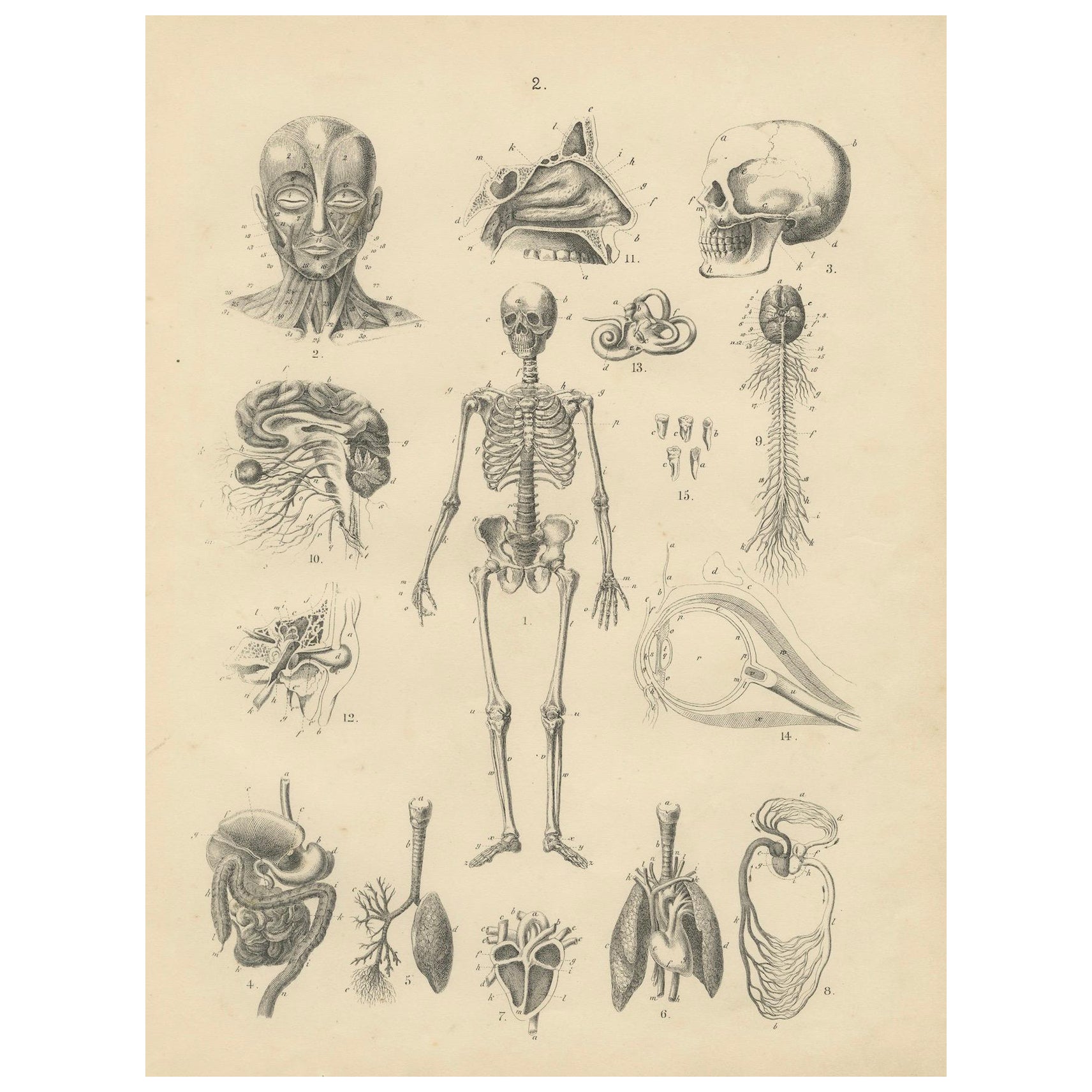
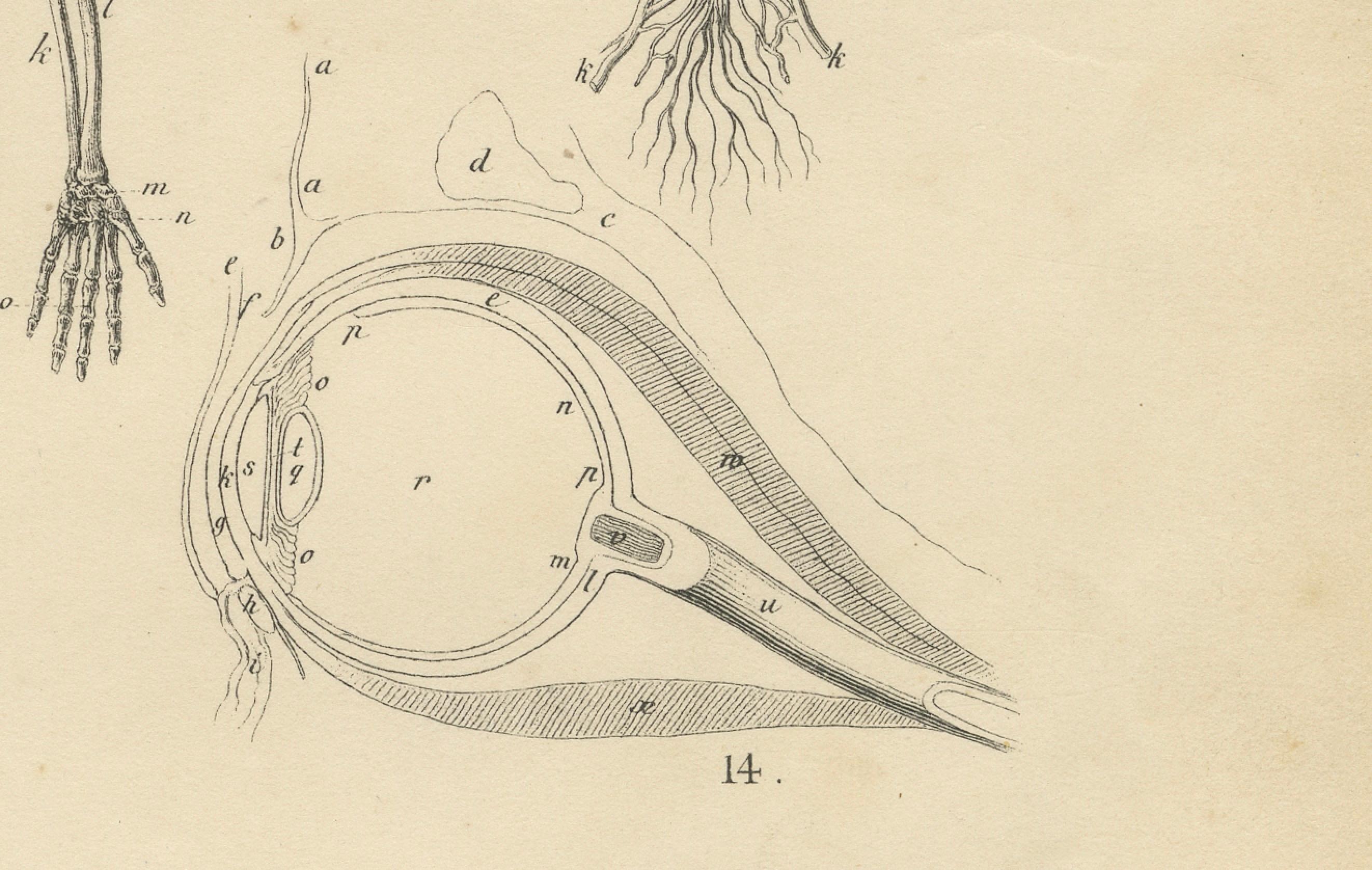
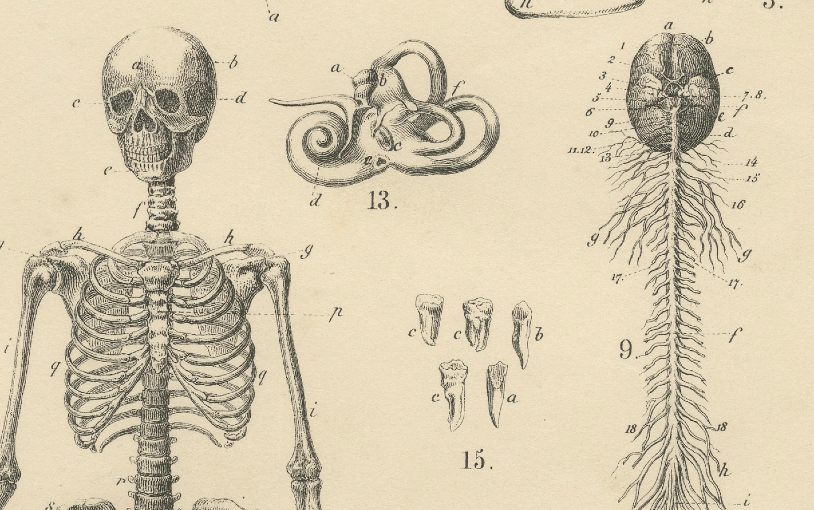
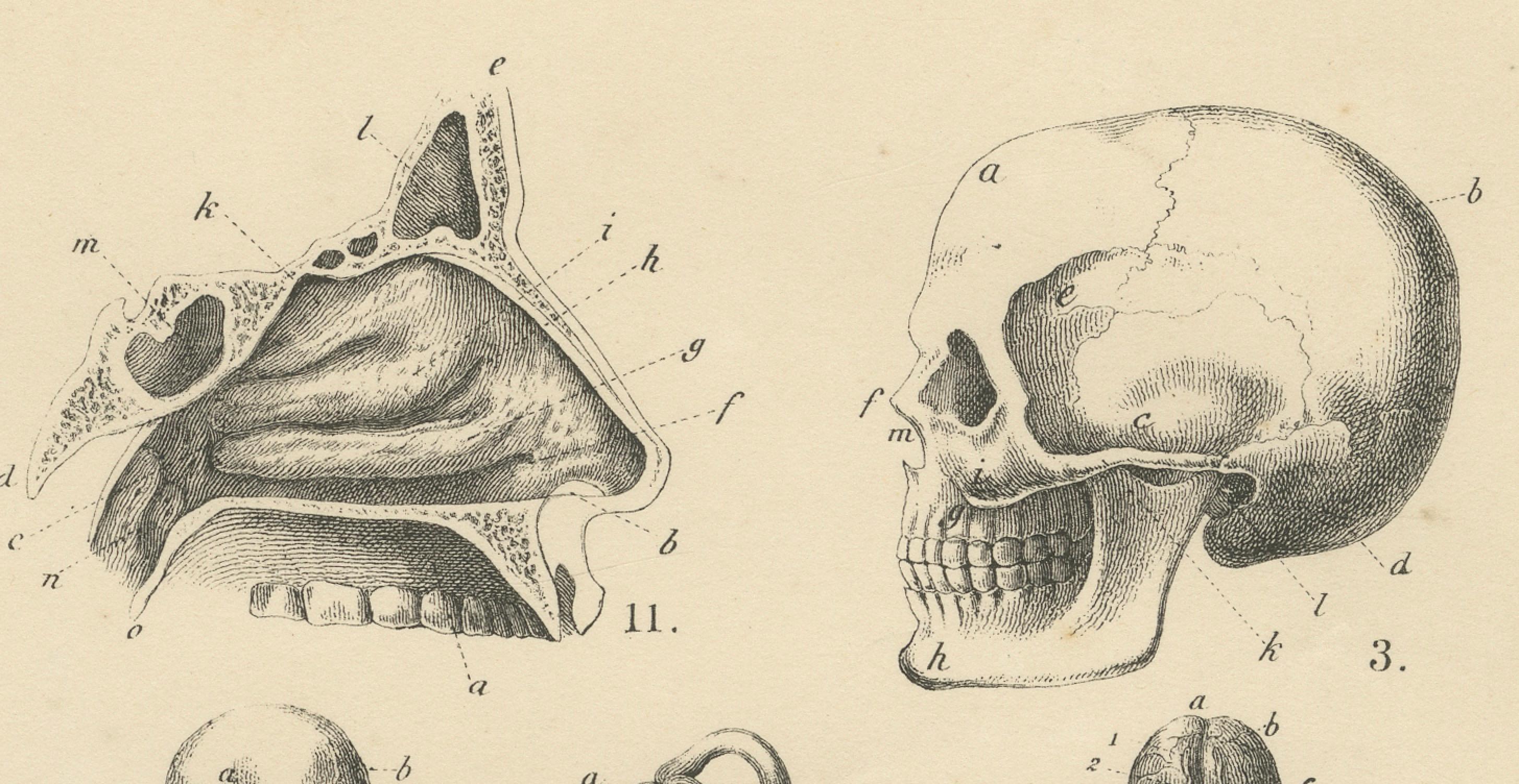
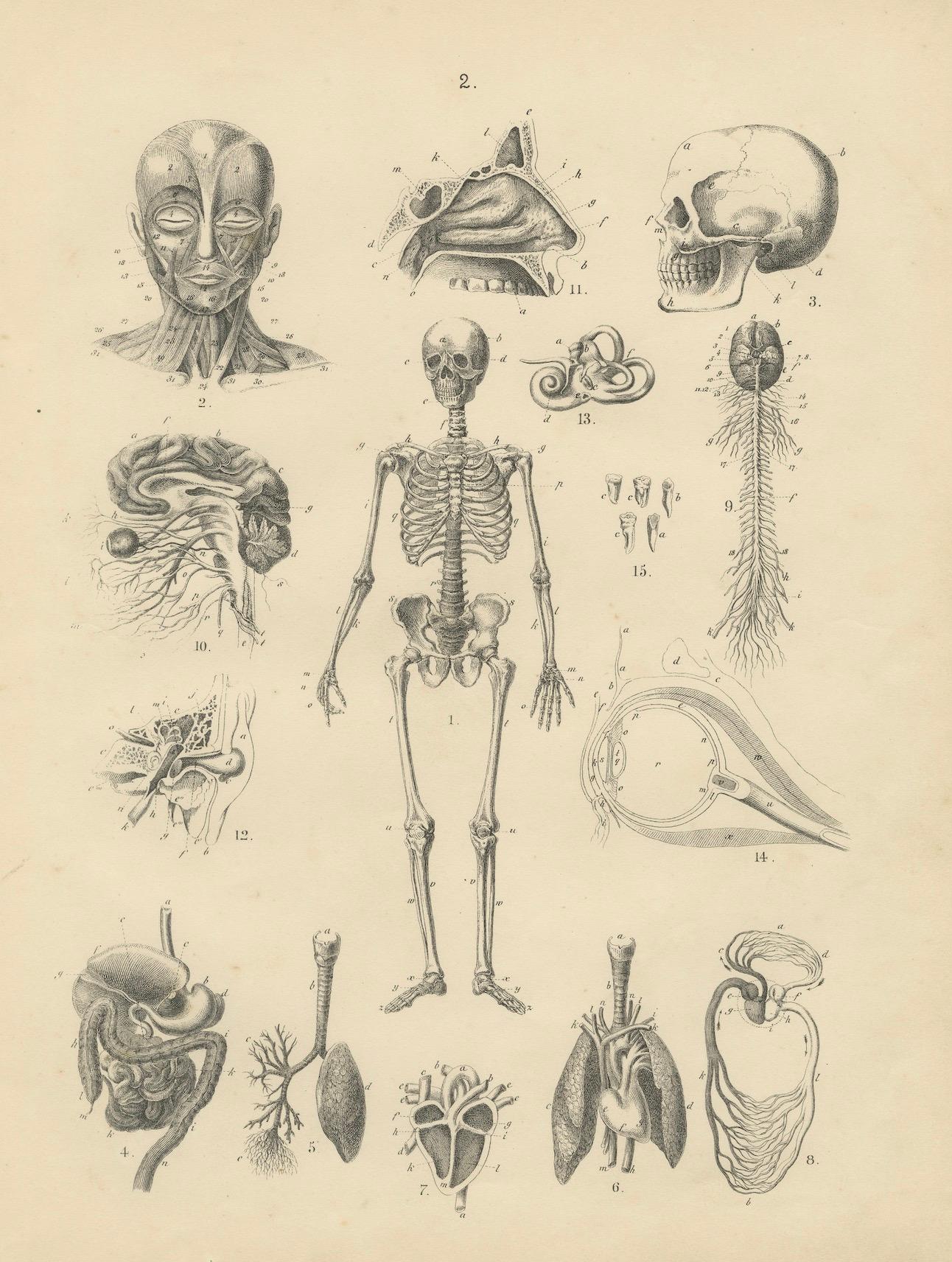
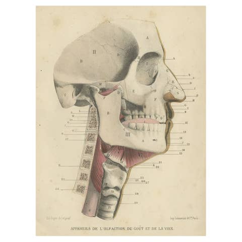
This detailed antique print presents a comprehensive anatomical study from the 19th century, depicting the human skeleton, skull, internal organs, nervous system, and other vital anatomical structures. It originates from the Atlas der Natuurlijke Geschiedenis voor scholen en huisgezinnen, adapted into Dutch by T.C. Winkler after Traugott Bromme, and published in Amsterdam by J.C. Loman in 1867. Intended as an educational tool, this engraving provided students and families with clear, labelled illustrations for learning human anatomy in a structured and accessible manner.
The labelled figures on this plate include:
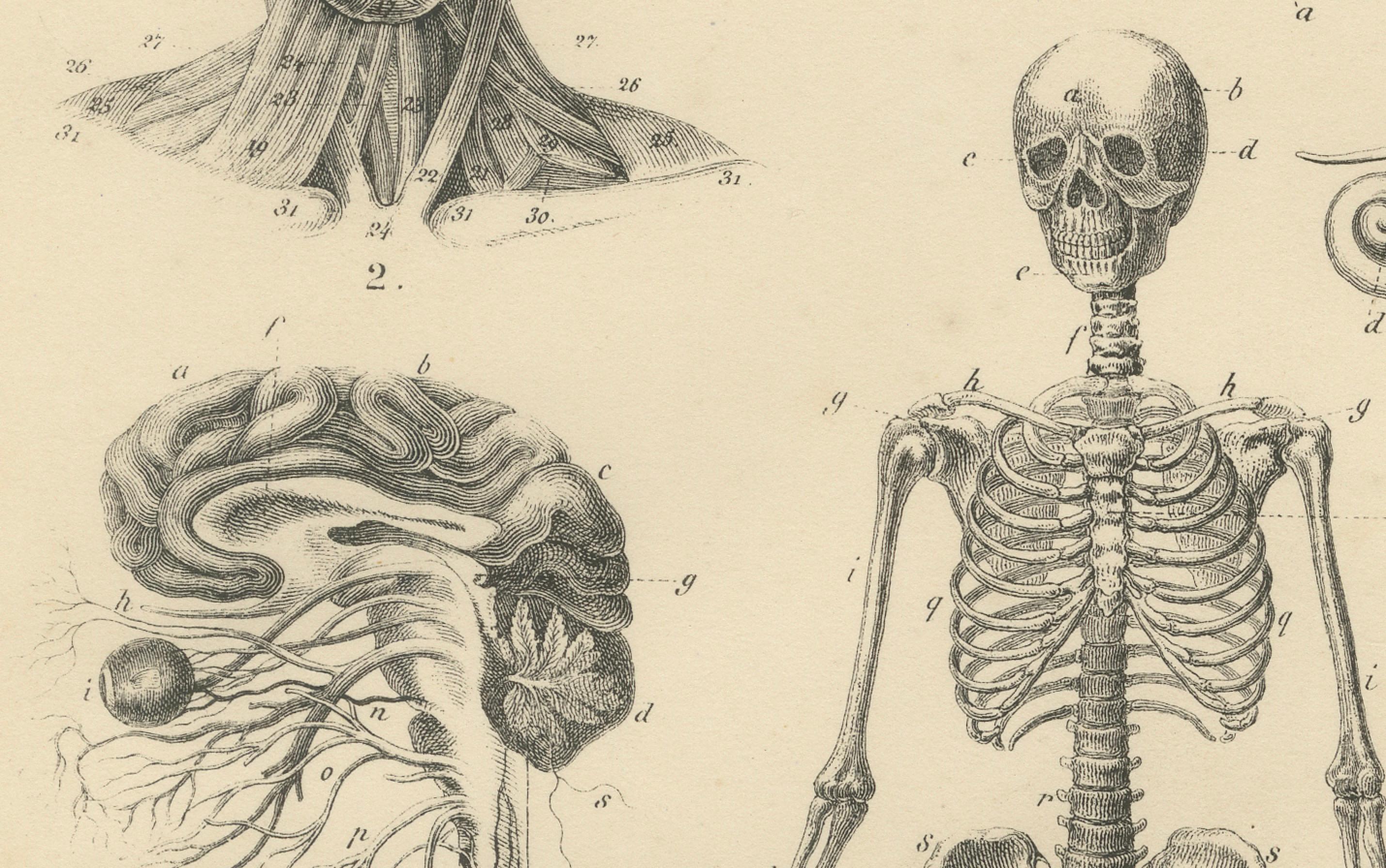
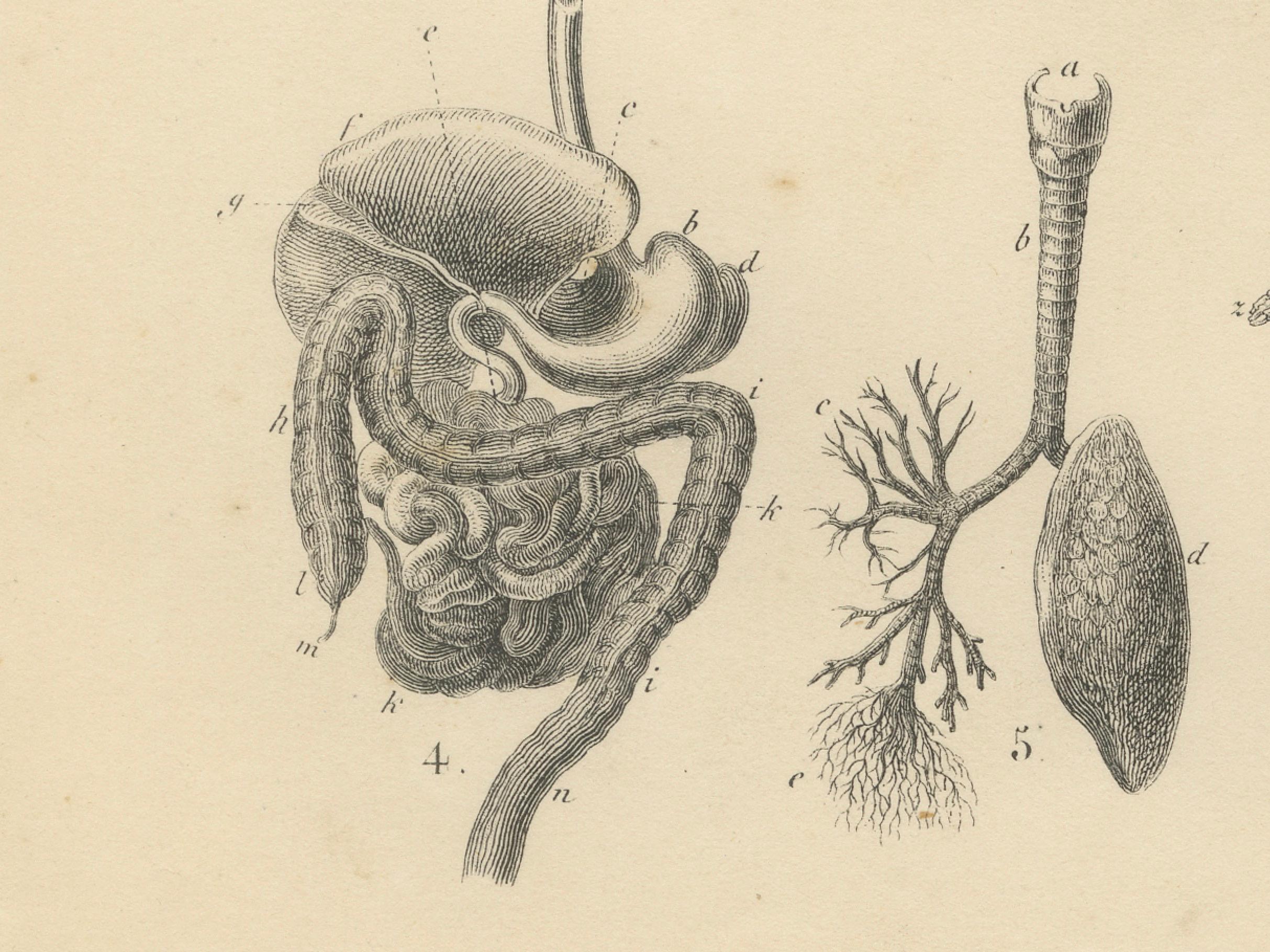
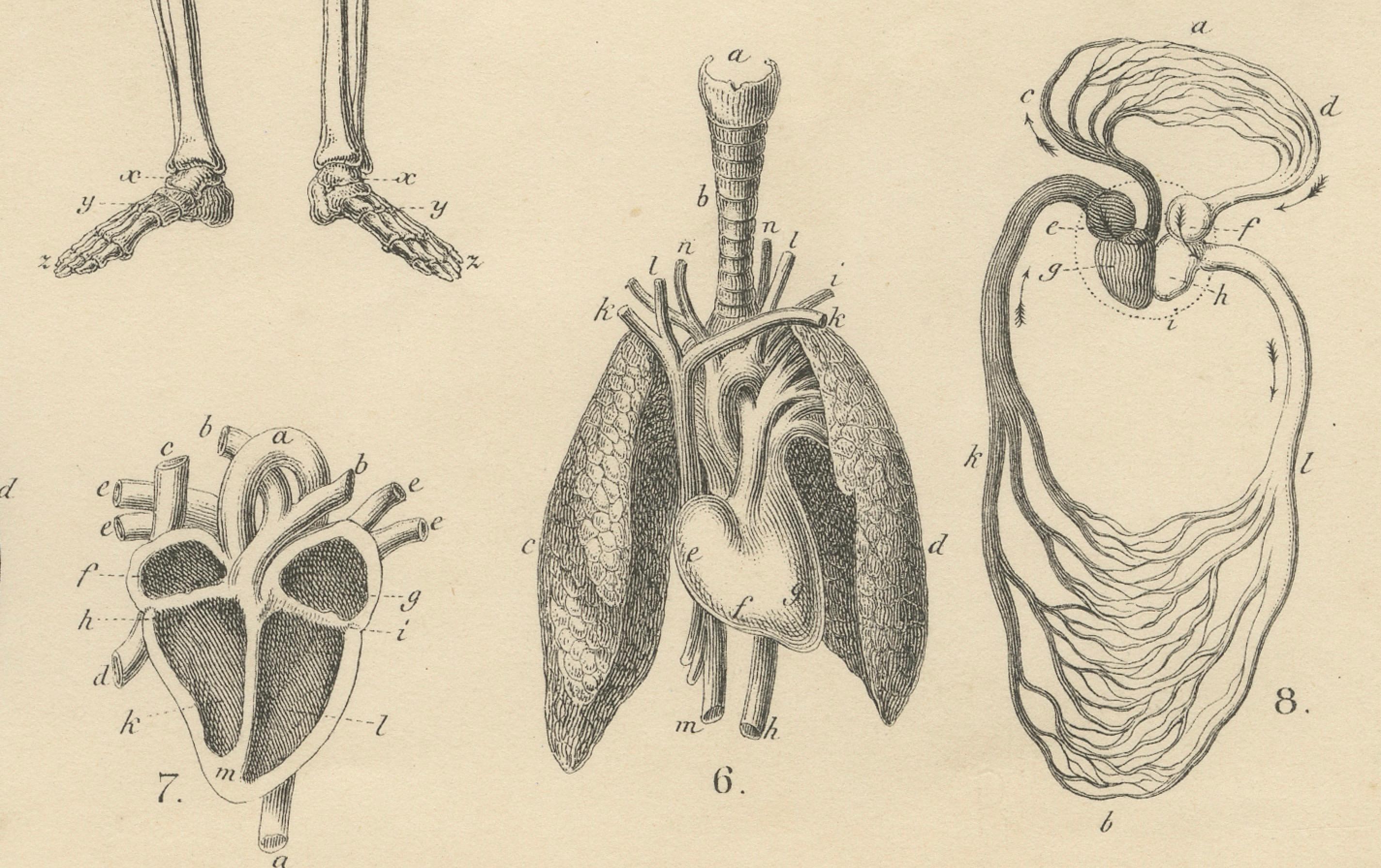
1. Full human skeleton 2. Facial muscles anatomy 3. Skull (side view) 4. Digestive system including stomach and intestines 5. Liver and lungs 6. Heart and lungs anatomy 7. Detailed heart anatomy 8. Urinary and reproductive system 9. Brain and spinal cord 10. Brain cross section with nerves 11. Nasal cavity anatomy 12. Ear structure with inner ear detail 13. Inner ear labyrinth 14. Eye anatomy with cross section 15. Teeth structure
The print was produced using fine copper engraving techniques, which allowed intricate anatomical lines and precise labelling essential for education. Unlike later chromolithographs, this print is uncoloured, providing clear contrast and scientific clarity typical for mid-19th century academic plates.
Such illustrations were vital teaching aids in schools, universities, and private homes, serving as reference tools for biology and medical students while also decorating classrooms and libraries with scientifically informative imagery. Today, they offer a window into the history of anatomical education and are valued for their clarity, precision, and decorative academic appeal.
Condition summary: Good condition for its age, with light toning and scattered small foxing spots mainly in the margins. Paper remains strong with no tears or repairs, and the engraving lines are crisp and well-preserved.
Framing tips: Frame this print in a black, dark walnut, or oak wooden frame combined with a light cream or white mount to enhance its scientific aesthetic. A thin gold inner border can add a subtle classical touch. Using UV-protective glass is recommended to preserve the paper and maintain its crisp engraved lines. This antique anatomy print is ideal for display in medical offices, libraries, or academic interiors, adding intellectual depth and historical character to any space.